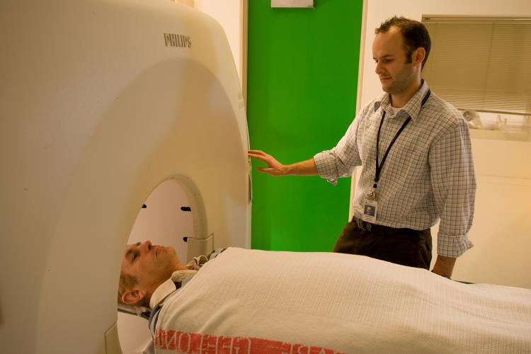
Being able to visualise the pathology linked to the disease is a big benefit for diagnosis and patient management.

Researchers from an IMI project that set out to get definitive data on the role of amyloid PET scans in Alzheimer’s disease diagnosis are hearing from physicians that being able to visualise the underlying pathology linked to the disease is a big benefit for diagnosis and patient management.
The ability to show patients the presence of amyloid plaque in the brain allows physicians to have ‘more meaningful’ conversations with their patients. Alzheimer’s disease is currently diagnosed based on cognitive symptoms, a clinical examination and a combination of brain scans such as CT or MRI which show structural abnormalities. Amyloid PET (positron emission tomography) scans are a newer technology and researchers from the AMYPAD project are trying to understand in which circumstances these PET scans are best deployed. They also want to find out at what stage in the disease they are most beneficial, and how an early PET scan might lead to a change in patient management.
“The hypothesis we are testing in the clinical study is that an early scan leads to a higher confidence in the diagnosis of the patient."
An ongoing clinical trial is examining how the PET scan influences the diagnosis of patients with cognitive complaints. Patients who come into memory clinicals are being categorised into three groups: those with very early complaints, called subjective cognitive decliners, those in an intermediate phase, called mild cognitive impairment and those with possible Alzheimer’s disease. Over 600 of the expected 900 patients have already been recruited at 8 sites across Europe. According to Gill Farrar, AMYPAD project leader, “The hypothesis we are testing in the clinical study is that an early scan leads to a higher confidence in the diagnosis of the patient. Physicians involved in the trials are also finding that it is useful to use the images to show the patient the presence or absence of pathology in the brain”
“There is also a lot of research going on in terms of how you articulate what you say to patients, as in how you share the information with respect to their increased risk of cognitive decline if the pathology is present. And one of the goals of this project is to work with the very talented physicians who are very experienced in this field to generate language on how best to share that knowledge.”
Video: what is the IMI AMYPAD project about?
Recruitment of the study should be completed in 2020, with results expected in 2021. The researchers have already started analysing the amyloid scans from these patients and hope to present some preliminary data next summer.
The recent news from Biogen with the projected submission of aducanumab to the FDA in the USA will be the first potential new drug for Alzheimer’s disease in many years and has initiated renewed interest in the use of pathology markers in the dementia field.
Gill Farrar is Global Medical Leader, PET Neurology and Digital Projects, Pharmaceutical Diagnostics GE Healthcare. She co-leads the IMI AMYPAD Project with Professor Frederik Barkhof. Professor Giovanni Frisoni is the Clinical Leader for the AMYPAD Diagnostic and Patient Management Study.
Read more
How competition nearly killed Alzheimer's research
Alzheimer's researchers use AI to get access to brains
Alzheimer’s project EPAD releases first wave of data to research community
